Showing posts with label pictures. Show all posts

Monday, November 22, 2021

Pictures Of Foot Cancer

Share on Pinterest A person may not notice symptoms of foot melanoma until the cancer has reached the later stages. Other common causes of skin cancer on your feet include.
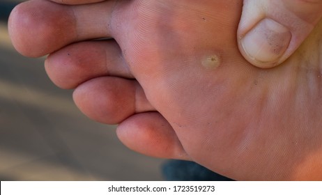
Foot Cancer Images Stock Photos Vectors Shutterstock

Melanoma is known as the most serious and deadly form of skin cancer.

Pictures of foot cancer. The earlier a skin cancer is diagnosed the easier it is to treat. You can also get invasive squamous cell carcinoma in chronic ulcers or scars after a burn or after radiotherapy. Find foot cancer stock images in HD and millions of other royalty-free stock photos illustrations and vectors in the Shutterstock collection.

It always involves a tendon sheath. Skin cancers can look very different. Download Bone cancer stock photos.

Lentigo Maligna Melanoma Melanoma. Giant cell tumor. The following picture shows you where to look.

Check the moles on your feet. 2135 dog cancer stock photos vectors and illustrations are available royalty-free. Affordable and search from millions of royalty free images photos and vectors.

Its not all that common on the feet and it is not a very aggressive form of cancer. The spot on the bottom of Paschals foot grew from a dot to a large lesion. Pay close attention to places on your feet that have been injured.

Previous Next 1 of 6 Melanoma pictures for self-examination. Research has shown that a foot. Search for dog cancer in these.

The American Academy of Dermatology advises that you watch skin spots for these features. Photos of skin cancer. Courtesy UK Athletics Skin cancer on the sole of the foot is more aggressive.

Melanin is the pigment that gives your skin color. It is important that all these symptoms are identified at an early stage so to. Itchy crusty or bleeding.

Foot Skin Cancer Causes and Symptoms. Browse foot cancer pictures photos images GIFs and videos on Photobucket. Melanoma a serious form of skin cancer is often curable if you find it early.

Even if the injury was years ago examine the area carefully. Squamous Cell Carcinomas A squamous cell carcinoma is the most common type of cancer of the feet. Skin cancer consists of tumors that grow in your skin and can eventually spread if left untreated.

See dog cancer stock video clips. Squamous cell carcinoma is most common in the face neck on the scalp or top side of the hands. A red or dark patch.

By thoroughly checking your feet you can find melanoma early. It often appears as a painful firm irregular lump in adults aged 20 to 50. 38 photos that could save your life.

They often appear as white or cream colored bumps on the skin and may ooze from time to time. The skin cancer pictures below show that melanomas can appear in an area that has not had much UV exposure in a persons lifetime such as the sole of the foot. Try these curated collections.

These melanoma pictures can help you determine what to look for. These precancerous lesions like the ones shown above on the back of a hand can turn cancerous. Lumps swellings fractures joint tenderness and pain are some common symptoms of bone cancer in ankle and foot.

So its important you visit your GP as soon as possible if you notice a change in your skin. As we noted above skin cancer of the foot can be caused by harmful and prolonged exposure to UV rays but thats not the only way cancers can develop. Heres a closer look at how skin cancer on your feet is caused diagnosed and treated.

It always involves a tendon sheath. A giant cell tumor may be found on the toes anywhere on the surface of the foot or deep inside the foot. Thousands of new high-quality pictures added every day.

Cancer in dogs anesthesia pet dog tumor dog recovery veterinarian anesthesia emergency room dog vet emergency dog in clinic labrador hospital gloves in vet. This type of skin cancer develops in the melanocytes the cells that produce melanin. The cancer can also look like an irritated wart that is painful to the touch.

A spot or sore.

Saturday, October 16, 2021

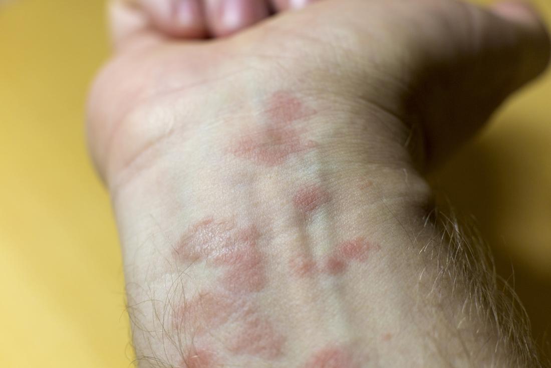
Itchy Rash On Wrist Pictures

Pictures and Diagnosis of Parasitic Rashes Itchy rash for 5 weeks Scabies. Dont delay your care at Mayo Clinic.

What Is This Rash On My Wrist

Heres a list of 21 with pictures.

Itchy rash on wrist pictures. Lichen planus is a skin condition characterized by small shiny reddish bumps. These rashes are usually caused by some kind of mite. What do autoimmune rashes look like.

10 common rashes on kids with photos. Typically contracted by prolonged contact with an infected individual. Common causes of a rash on the wrist include.

Notice a rash on your little one. 1 11 Photo. Autoimmune rashes look different depending on which autoimmune condition is causing the skin rash.

A wrist rash refers to change in the skin which affects the color appearance or textures of the skin. It is also possible for this kind of wrist accessories to cause itchy wrists without the involvement of an allergic reaction. There are many different causes for rashes.

It starts as a red rash such as the one seen in the shingles photo above but will turn into fluid-filled blisters. By Todays Parent May 1 2018. It can appear hours to days after exposure.

Allergens usually cause a shiny blistered itchy rash while irritants tend to cause a dry scaly less itchy rash. Scalp psoriasis may cause plaque buildup that results in hair loss. While the shingles rash typically develops on the torso first it can spread to almost any other place on your body.

See pictures of distinct types of COVID-19 rashes later in this article. Keep reading for more on four of the most common wrist rashes. See pictures of common skin rashes that can occur due to a variety of factors including infections heat allergens and medications.

Sometimes these are punctuated by. This article gives you a breakdown on the causes of both itchy and non-itchy wrist bumps symptoms and images pictures of how these bumps look like the treatments offered for wrist bumps and the common home remedies for wrist rash. It does not produce symptoms until the host becomes allergic to the mites after around 3 weeks.

An itchy rash on both wrists could be an indication of an allergic reaction to wristbands sweater or shirt cuffs etc. These rashes can present quite early on in the infection but can also last a long time afterwards. For example cutaneous lupus may cause a scaly red patch that does not hurt or itch.

Read on for pictures of common rashes that occur on the wrist along with their symptoms causes and treatments. Extremely itchy rash may be pimply made up of tiny blisters or scaly. A common scenario is where wrist accessories are worn too tight or over an extended period of time.

See pictures of common skin rashes that can occur due to a variety of factors including infections heat allergens and medications. Lichen planus may cause purplish itchy flat bumps on. Heres help figuring out common rashes on kids and what to do about them.

Sunday, September 6, 2020

Laboratory Glassware Names And Pictures Pdf

What would a chemistry lab be without glassware. Chemistry Laboratory Glassware Chemistry Demonstration.

Design Elements Laboratory Equipment Laboratory Equipment Vector Stencils Library Chemistry Laboratory Glassware

COMMON LABORATORY TECHNIQUES In all volumetric glassware pipet buret volumetric flasks graduated cylinder etc it is necessary to read the level of a liquid.

Laboratory glassware names and pictures pdf. Chemistry Lab Glassware List With Image. Let us see their features and uses in detail. Laboratory Glassware List Pdf.

Use Of Laboratory Glassware Pdf. Laboratory Glasswares And Their Uses Pdf. Laboratory Glass Wares And Their Uses With Pictures Pdf.

Lab Equipment Glassware List. Various equipment in the organic chemistry lab. Corning Life Sciences provides certificates for certifiedserialized PYREX items that are calibrated in our ISOIEC 17025-accredited laboratory.

Usually this meniscus curves downward to a minimum at the center. Laboratory glassware refers to a variety of equipment used in scientific work and traditionally made of glassGlass can be blown bent cut molded and formed into many sizes and shapes and is therefore common in chemistry biology and analytical laboratoriesMany laboratories have training programs to demonstrate how glassware is used and to alert firsttime users to the safety hazards. July 10 2019 8541.

Weighing paper used for weighing and transferring powers and crystals wire gauze used to support a container such as a beaker or flask during heating. A liquid in a small-diameter container will form a meniscus or curve at the surface of the liquid. The indication DN in the following table refers to the nominal size of the laboratory flat flange.

Glassware used in microscale work. Find laboratory glassware stock images in HD and millions of other royalty-free stock photos illustrations and vectors in the Shutterstock collection. This is a plain and simple cylindrical trough made of glass.

George Doyle Getty Images. Digital Certificates Will Improve Convenience and Reduce Paper Waste. On request O-rings made of silicone or Viton are available.

Glassware commonly used in organic chemistry for conducting reactions and purifications. Print Lab equipment names and pictures Quizlet Quizlet. About Ace Glass Ace Glass Incorporated founded 1936 in Vineland NJ is a leader and innovator in the manufacture of premium scientific glassware lab equipment and glass apparatus.

An erlenmeyer flask is a cone-shaped container with neck so you can hold the flask or attach a clamp or use a stopper. Test tube for chemical laboratory or science lab medicine or pharmacology liquid fluid measurement. Name Picture Use Ring stand Supports the bunsen burner iron ring pipestem triangle and other items often while heating a substance.

Chemistry Glassware Names and Uses. Select from premium Laboratory Glassware of the highest quality. Tools used in all chemistry courses.

Glassware used in all chemistry courses. Be able to identify the name of each piece of equipment as well as its function or use in the laboratory. Effective January 1 2020 customers will be able to download digital certificates as PDF files from the Corning Life Sciences online resource library simply by entering the item ID number and.

Lab glassware used in most labs. Glassware In Chemistry Lab Pdf. Each of these containers has its own unique form and purpose.

There can be a large amount of glassware in a lab. Clamps for securing apparatuses. There are several sizes that can be chosen from based on the amount of liquid that needs to go through them quickly.

Set of isolated glassware flask or glass bottle for chemistry on transparent background. Over our long history thousands upon thousands of scientific papers research and discoveries have been attributed to Ace Glass. They can be made of plastic or glass and can have either a short stem or a long stem depending on what they are needed for.

Find the perfect Laboratory Glassware stock photos and editorial news pictures from Getty Images. It is transparent and has a flat bottom. Chemistry Glassware Names and Uses.

Front view laboratory glassware stock pictures royalty-free photos images. A lab funnel is just like any other funnel except that it was designed to be used in a laboratory setting. Common types of glassware include beakers flasks pipettes and test tubes.

Biology Set of isolated glassware flask or glass bottle for chemistry on. Glassware Laboratory Apparatus And Their Uses Pdf. LABORATORY GLASSWARE ACCESSORIES FOR LABORATORY FLAT FLANGES O-ring seals These O-rings made of silicone and FEP-lined have a very high elasticity ensuring a good recovery and re-use.

Laboratory Equipment and Functions1 of 5 Study the table below. Thousands of new high-quality pictures. Laboratory glassware watch glass to hold solids while being weighed or as a cover for a beaker.

Monday, July 29, 2019

Urinary Tract Infection Pictures

It is characterized by painful and frequent urination cloudy urine and a fever with back pain among other symptoms and is caused by a growth of bacteria in the urinary tract. Signs of male urinary tract infections include urinating more frequently than normal as well as pain when urinating.

Urinary Tract Infection Prevention

Intercourse and ejaculation may be painful as well.

Urinary tract infection pictures. The sufferer may feel more tired than usual or be passing urine that has a milky or cloudy appearance. Pseudomonas in urine may occur as a result of sexual activity. The ureters are the tubes.

Thousands of new high-quality pictures. Browse 732 urinary tract infection stock photos and images available or search for cystitis or urinary system to find more great stock photos and pictures. A urinary tract infection UTI is an infection involving the kidneys ureters bladder or urethra.

Urinary tract infection stock pictures royalty-free photos images Vector medical poster symptoms of cystitis. A UTI or urinary tract infection is a common problem among many women. Try these curated collections.

Coli urinary tract infection cell biology stock photos and images available or start a new search to explore more stock photos and images. - urinary tract infection stock pictures royalty-free photos images. The patient may need an evaluation in the future to check for a recurrence a potential concern with urinary tract infections.

Affordable and search from millions of royalty free images photos and vectors. Find urinary tract infection stock images in HD and millions of other royalty-free stock photos illustrations and vectors in the Shutterstock collection. Download in under 30 seconds.

Browse 746 urinary tract infection stock photos and images available or search for bladder or urinary system to find more great stock photos and pictures. Getty Images UTIs in dogs are most often caused by bacteria that enters through the urethral opening and moves upward into the urinary. The relatively short length of the urethra in women increases the chances of.

Having issues down there. Picture of the urinary tract. Download Urinary tract infection images and photos.

Photos of the Bacteria in Urinary Tract Infections. Causes Of Urinary Tract Infections In Dogs Picture Credit. Your Urinary Tract Infection stock images are ready.

Medical form with diagnosis Urinary tract infection. Female urologyurinary tract lungsprostate 3drenal diseaseadrenal glandsexcretory systemadrenal glandvasectomy. Dreamstime is the worlds largest stock photography community.

These are the structures that urine passes through before being eliminated from the body. February 11 2021 Cranberry pills can be used to treat a UTI. Use them in commercial designs under lifetime perpetual worldwide rights.

A urinary tract infection UTI occurs when germs bacteria invade the urinary tract. Warning some of the photos are extremely graphic in nature. 897 male urinary tract stock photos vectors and illustrations are available royalty-free.

Diseases of the genitourinary system. The kidneys are a pair of small organs that lie on either side of the spine at about waist level. Download all free or royalty-free photos and vectors.

Bacteria can travel from the feces to the urethra particularly in women. Male urinary tract images. The urinary tract is made up of your kidneys bladder ureters and urethra.

See male urinary tract stock video clips. Download urinary tract infection stock photos. Below is a collection of pictures of the effects caused by Urinary Tract Infections UTI.

Urinary Tract Infection UTI Pictures. Men who have experienced kidney stones are prone to urinary tract infections. Over 425 Urinary tract infection pictures to choose from with no signup needed.

Monday, December 31, 2018

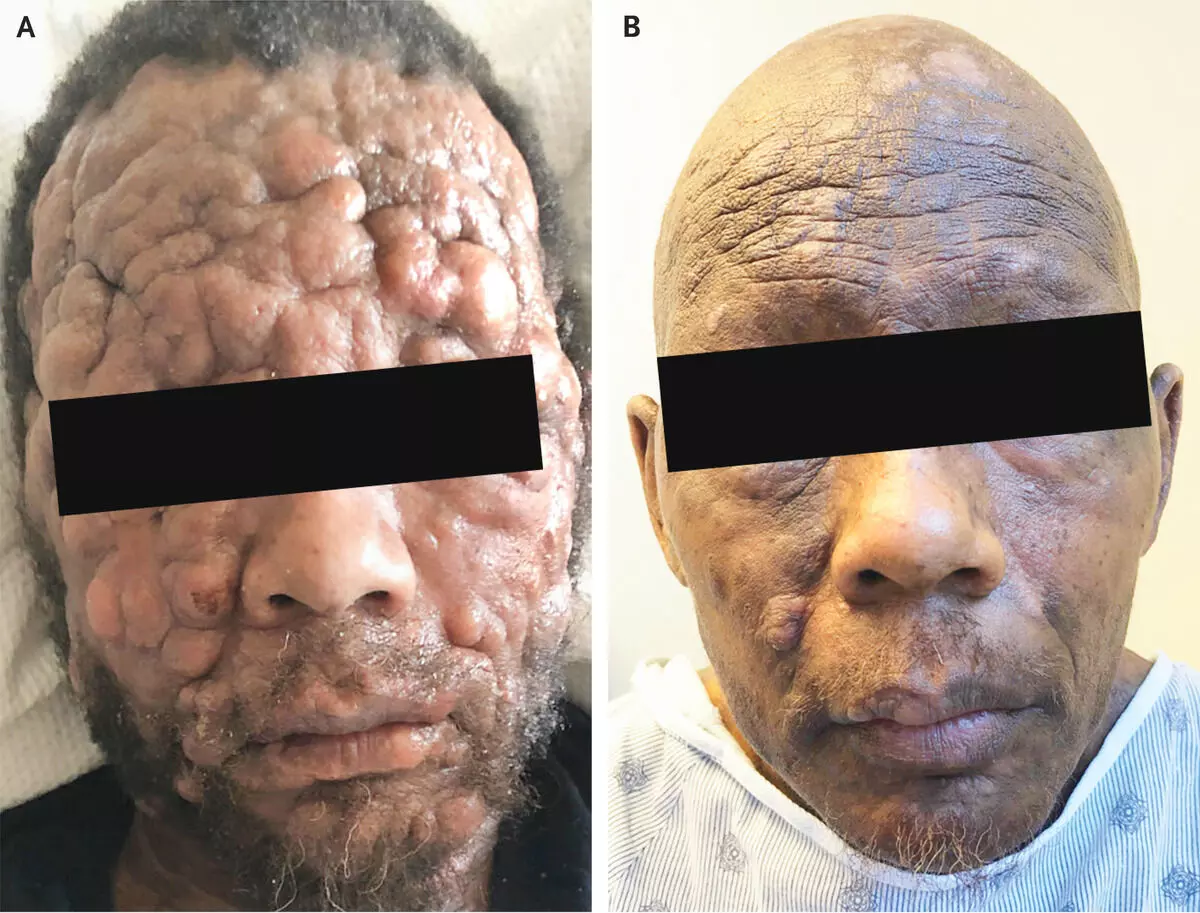
Mycosis Fungoides Pictures

Skin candida fungal infection on skin man fungus athlete care nail infection feet man woman nails dermatophytes ringworm of the body onychomycosis tinea. Find mycosis fungoides stock images in HD and millions of other royalty-free stock photos illustrations and vectors in the Shutterstock collection.

Mycosis Fungoides Causes Symptoms Diagnosis Treatment

Mycosis fungoides also known as Alibert-Bazin syndrome or granuloma fungoides is the most common form of cutaneous T-cell lymphoma.

Mycosis fungoides pictures. Bereits mit dem bloßen Auge kann der Hautarzt Veränderungen erkennen CorbisBoissonet Übersicht. Ad Emuaid Kills Toe Nail Fungus on Contact and Eliminates 9999 of Bacteria in 1 Minute. Symptoms include rash tumors skin lesions and itchy skin.

Wer an einer Mycosis fungoides leidet. What is Mycosis Fungoides. Ad Emuaid Kills Toe Nail Fungus on Contact and Eliminates 9999 of Bacteria in 1 Minute.

It is very difficult to determine if somebody will have fungoides of the mycosis diverse biopsies is required generally on a number of years before the diagnosis can be made. This condition is one of the most common types of T-cell lymphoma. Try these curated collections.

Tumour stage mycosis fungoides. Thousands of new high-quality pictures added every day. It can spread to other parts of your body including the internal organs and blood with different degrees of severity.

While the cause remains unclear most cases are not hereditary. Browse 3 mycosis fungoides stock photos and images available or search for lymphoma or cd30 to find more great stock photos and pictures. Anaplastic large cell lymphoma.

From left Sharon Cohen Biotechnology Industry Organization Wendy Andrews University Medical Center in. Mycosis Fungoides - Pictures Staging Symptoms Treatment and Causes. The patches or plaques can look like eczema psoriasis or another common skin problem.

It generally affects the skin but may progress internally over time. Although the terms MF and CTCL are often used interchangeably this can be a source of confusion. It can be hard for your doctor to know for sure that you have mycosis fungoides.

All cases of MF are CTCL but not all CTCL cases are MF. January 30 2021 Mycosis fungoides is one of many types of lymphoma. Search for mycosis in these categories.

This medical condition is one type of lymphoma and will often infect the skin. End The Suffering Of Toenail Fungus In Days. Mycosis fungoides follows a slow chronic indolent course and very often does not spread beyond the skin.

Mycosis fungoides and the Sezary syndrome is a disease in which certain cells of the lymph system. Mycosis fungoides is characterized by a scaly red rash that develops on the skin particularly on areas that are not usually exposed to the sun. Mycosis fungoides is a disease in which T-cell lymphocytes a type of white blood cell become malignant cancerous and affect the skin.

See mycosis stock video clips. This results in one rash. It often manifests first as a simple red rash.

A lymphoma rash such as mycosis fungoides MF can be easy to confuse with other skin conditions such as psoriasis or eczema which can cause similar symptoms. A sign of mycosis fungoides is a red rash on the skin. In this condition white blood cells called lymphocytes begin to attack the skin.

In fungoides of the mycosis lymphoma of the blood remains in the biggest part in the skin. Ulcerated tumour stage mycosis fungoides. 2738 mycosis stock photos vectors and illustrations are available royalty-free.

End The Suffering Of Toenail Fungus In Days. Mycosis fungoides MF is the most common form of cutaneous T-cell lymphoma CTCL. Mycosis fungoides is a type of lymphoma that effects the skin in a form of cancer called cutaneous T-cell lymphoma CTCL.

Most cases are in people over 20 years of age and it is more common. Mycosis fungoides and Sézary syndrome are types of cutaneous T-cell lymphoma. Mycosis fungoides and Sézary syndrome are diseases in which lymphocytes a type of white blood cell become malignant cancerous and affect the skin.

Monday, October 1, 2018

Necrotizing Fasciitis Pictures

Here are some pictures of Necrotizing Fasciitis. Halloween skulls Severed head burnt halloween skulls special effect makeup necrotizing fasciitis stock pictures royalty-free photos images.

Necrotizing Fasciitis A Plastic Surgeon S Perspective Sciencedirect

Necrotizing fasciitis is a dangerous infection of soft-tissue that starts in the subcutaneous tissue just below the skin and spreads along the flat layers of fibrous tissue that separate different layers of tissue fascial planes.

Necrotizing fasciitis pictures. Sponsored content Related information. Update to the Clinicians Challenge Award 2018 An update of our c ontinuing research into dermatological image recognition using deep learning and vision algorithms. The Necrotizing Fasciitis images will also help you understand the symptoms of the disease much better.

Brown and yellow leaf spots Necrotic dead spots with chloritic yellow halos on a tropical leaf necrotizing fasciitis stock pictures royalty-free photos images. Accurate diagnosis rapid antibiotic treatment and prompt surgery are important to stopping this infection. Fascial planes are bands of connective.

Go through these Necrotizing Fasciitis photos to see how the condition looks like. Symptoms of necrotising fasciitis usually come on quite quickly after the break in the skin occurred. Intravenous Drug Users are at Risk.

Necrotizing fasciitis is often described in the media as the flesh eating bug and this offers a fair description of what it involvesIt is an extremely serious infection because one in four of the people who develop it will die as a result. It most commonly occurs in the arms legs and abdominal wall. Fascia is part of connective tissue.

Necrotising fasciitis is a rare but serious bacterial infection that affects the tissue beneath the skin and surrounding muscles and organs fascia. Picture of Necrotizing Fasciitis. Septic shock would be usually one of the most fatal outcomes that could result from necrotizing fasciitis.

What are the symptoms of necrotising fasciitis. Necrotizing fasciitis NECK-re-tie-zing FASH-e-i-tis is a rare bacterial infection that spreads quickly in the body and can cause death. The infection may spread very quickly and even be fatal.

You can also find pictures of Necklace of Venus Oculocutaneous albinism Ochronosis. At first the affected skin looks slightly. It then tends to develop very rapidly over a few days or even less.

Find necrotizing fasciitis stock images in HD and millions of other royalty-free stock photos illustrations and vectors in the Shutterstock collection. Necrotizing fasciitis is a relatively rare condition that intravenous drug users are at risk of developing. Necrotizing fasciitis can be caused by several different types of bacteria and the infection can arise suddenly and spread quicklyEarly signs include flu-like symptoms and redness and pain around the infection site.

Thousands of new high-quality pictures added every day. Its sometimes called the flesh-eating disease although the bacteria that cause it do not eat flesh but release toxins that damage nearby tissue. 2 Orthopeadics and Traumatology Department Geneva University Hospital Geneva Switzerland.

Necrotising Fasciitis of the Hand. Click here for Necrotizing fasciitis pictures. Necrotizing fasciitis is a serious infection of the skin the tissue just beneath the skin subcutaneous tissue and the tissue that covers internal organs fascia.

1 Hand Unit Geneva University Hospital Rue Grabielle Perret Gentil 4 1211 Geneva Switzerland. Picture of Necrotizing Fasciitis. See smartphone apps to check your skin.

If left totally untreated mortality rates could reach to as much as 73. Necrotizing fasciitis sometimes referred to by the name as flesh-eating bacteria is a condition in which bacteria infect and destroy the skin subcutaneous fat and fascia. Necrotizing fasciitis is a term that describes a disease condition of rapidly spreading infection usually located in fascial planes of connective tissue that results in tissue necrosis dead andor damaged tissue.

Highlights Seek And Find

Thousands of new high-quality pictures added every day. HIGHLIGHTS KIDS Watch More On YouTube. Birds Of A Feather Highlights Kids Create...