4th intercostal space right sternal border. Left leg limb lead is red and is.
 3 Lead Placement Respiratory Therapy Nicu Nurse School Motivation
Additional Lead placements Right sided ECG electrode placement.

3 lead ecg placement. It is positioned in the 5th Intercostal Space on the Left Mid-Clavicular Line the vertical line which passes through the center of the Clavicle Eldrige et al. A red lead is placed on the right wrist or shoulder- known as AVr. Einthovens Triangle is formed when when the three ECG leads are placed on the patient.
5 Instruct the patient to relax in a supine position. Deviation of lead placement even by 20-25mm from the correct position can create clinically significant changes on the ECG. 4 Attach the ECG clips from the patient cable to the electrodes according to color coding.
ECG electrode placement is standardised allowing for the recording of an accurate trace but also ensuring comparability between records taken at different times. Poor electrode placement can result in mistaken interpretation and then possible misdiagnosis patient mismanagement and inappropriate procedures Khunti 2013. Enter Technician Physician and.
3 Adhere electrodes to the patient. 3 and 5-lead monitoring take electrodes that are used for the limbs in 12-lead monitoring and instead place them on the chest wall in order to reduce artifacts ECG signals that are from sources other than the heart caused by patient movement Khan 2015. Chest Precordial Lead Placement.
The three leads used in a 3-lead ECG are made up of foam electrodes with an adhesive side that attaches to the skin like an adhesive bandage. As a result torso leads are preferable as they allow for better quality ECGs. An ECG lead is a graphical description of the electrical activity of the heart and it is.
YT and other entities do not have any copyrightsBeep sound is protected by copyrig. Midway between leads V2 and V4. 3-Lead ECG A 3-Lead ECG uses 3 electrodes that are labeled white black and red.
5th intercostal space anterior axillary line. A 5-Lead ECG uses 4 limb leads and 1 chest lead. They are disposable intended to be thrown away after one use.
7 Select ECG button. Each lead is connected by a cord directly to an ECG machine or to a wireless device that sends the signal to an ECG monitor. 4th intercostal space left sternal border.
To opposite side of this card for proper placement b. The recommended 3-wire ECG lead placement is as follows. 3 lead ECG is non-diagnosticthus does not provide a clear view of the entire heart.
5th intercostal space midclavicular line. 3-leads ECG -EKG 3-odprowadzeniaIm owner of all material in this video. 6 Select New Test button.
Even though limb lead placement is often represented in terms of the apices of an equilateral triangle known as the Einthoven triangle Einthovens law is entirely independent of any assumptions about geometric placement of the 3 electrodes. Before discussing the ECG leads and various lead systems we need to clarify the difference between ECG leads and ECG electrodesAn electrode is a conductive pad that is attached to the skin and enables recording of electrical currents. These are the most common 3 lead ECG placements.
Place LA black electrode under left clavicle mid-clavicular line within the rib cage frame. V7 Left. Rub skin with alcohol wipes.
These 3 leads monitor rhythm monitoring but doesnt reveal sufficient information on ST elevation activity. A complete set of right-sided leads is obtained by placing leads V1-6 in a. These give you more views of the heart and can help inform your treatment plans.
The 3rd ECG chest lead to be placed is V4 C4. Instead it provides a basic view of the electrical pathway of the heart triangulated between the 3 leads. 3-wire Lead Placement AHA Place RA white electrode under right clavicle mid-clavicular line within the rib cage frame.
These colors are not universal as two coloring standards exist for the ECG discussed below. Erhardt et al first described the use of a right sided precordial lead CR 4 R or V 4 R in the. From electrodes to limb leads chest leads 12-lead ECG.
ECG limb lead placement diagram. As a consequence the 3 standard limb leads contain only 2 independent pieces of information. Right arm limb lead is white white goes to the right forearm proximal to the wrist.
At a minimum lead V4 should be placed on the 5th intercostal mid-clavicular exact opposite of the regular left side placement if an inferior infarct was originally seen in leads II III and AVF. Left arm limb lead is black and is considered the Earth lead and is placed at the forearm proximal to the wrist. V4R ECG lead placement.
Watch a video on ECG leadelectrode placement.
3 Lead Left And 12 Lead Right Ecg Electrode Placement Download Scientific Diagram
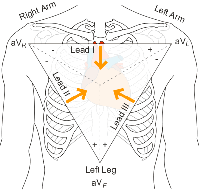 Bipolar Leads Ecg Lead Placement Normal Function Of The Heart Cardiology Teaching Package Practice Learning Division Of Nursing The University Of Nottingham
 Ecg Lead Positioning Litfl Ecg Library Basics
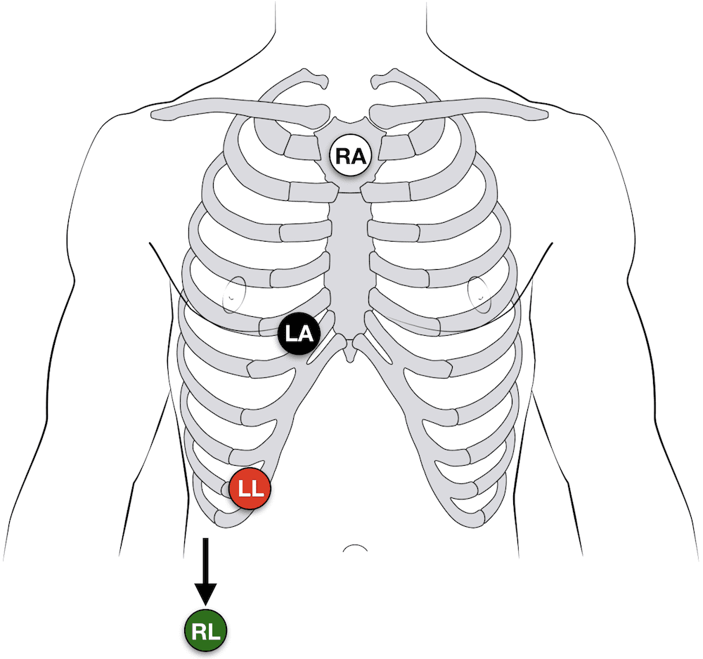 Ecg Lead Positioning Litfl Ecg Library Basics
 How To Place 3 Lead Ekg Youtube
Https Www Mindraynorthamerica Com Cmsadmin Uploads Ecg Lead Placement Proc 7664reva Pdf
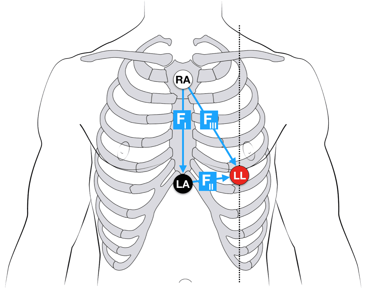 Ecg Lead Positioning Litfl Ecg Library Basics
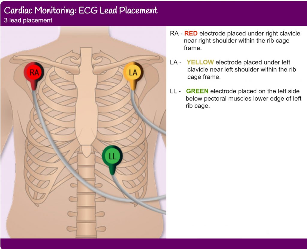 3 Leads Ecg Cable And Placement Yqf Medical Cable
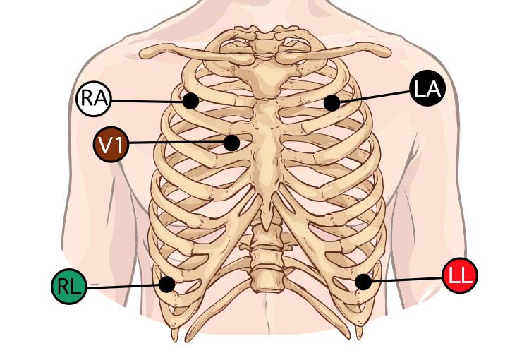 5 Lead Ecg Placement And Cardiac Monitoring Ausmed

 Figure 10 3 Lead Ecg Placement Options Image From The Hrw Project Ekg Leads Nursing Mnemonics Ekg Placement
 How To Place 3 Lead Ekg Youtube
 3 Lead Ecg Electrode Placement Diagram Google Search

No comments:
Post a Comment
Note: Only a member of this blog may post a comment.